
In 2020, while the world suffered from the worse pandemic in decades, the acronym PCR, became familiar to most of us. PCR (Polymerase Chain Reaction) procedure has recently been realized as a major tool specifically in diagnosing people contaminated by Coronavirus. PCR was invented in the early 80’s and became one of the most widely used methods in molecular biology. It is used mainly in labs to produce sufficient quantities of DNA or RNA from a sample where this material is in limited quantities.
The ultimate goal of PCR is to produce multiple identical DNA fragments from one single DNA or RNA strand. PCR is often used in multiple applications such as forensics, detection of microorganisms, prenatal diagnosis, medical diagnostics, and agriculture, to name a few. See the chart to the right for an example of reverse transcription polymerase chain reaction which is a laboratory technique combining reverse transcription of RNA into DNA, along with amplification of specific DNA targets using PCR.

Figure 1: Cycle of RT-PCR (reverse transcriptase PCR)
This contamination can come from the environment, or from cross contamination by other preamplified samples. Depending on the application, the impact of contaminations can be dramatic and could result in bad diagnostics or could compromise criminal investigations.
In order to optimize PCR and to avoid compromised results, the PCR process must be clearly organized, following 3 major steps, each of them requiring stringent protocols:
1 – Lysis of cells and extraction of DNA or RNA
2 – Preparation of PCR mixture before amplification
3 – Amplification by itself in a thermocycler
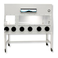
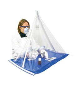
DNA and RNA are contained inside cells, and more generally, in the cell nucleus. Prior to PCR, the first step is the extraction of DNA or RNA from the cell. This can be done using different strategies. During this step, the sample must be protected to avoid it being contaminated by environmental pollution, while at the same time the operator must be protected against hazardous chemical that are used to extract DNA or RNA. If the sample is a pathogenic micro-organism, this part of the procedure must be performed in a Biological Safety Cabinet type 2. At the end of step one, the extracted sample must be isolated, and the next step must absolutely be performed in a second work station.
During step 2, also called pre-PCR, extracted samples are mixed with all reactants that are needed to perform amplification. These reactants include primers that hybridize into flanking sequences on opposing strands of the target, 4 deoxyribonucleoside triphosphates and a DNA polymerase along with buffer, co-factors of enzymes and water. During this sequence, the sample is highly sensible to both environmental and cross contaminations, as DNA or RNA strands are no longer protected by cells, and are in very limited quantities. Environmental contamination could come from dust air pollution that embeds micro-organisms or other biological material, and cross-contamination could come from a previous sample that was manipulated in the same enclosure.

The best way to protect the sample against air contamination, is to blow purified air on the sample. Air is purified by passing through high efficiency particulate filters such as HEPA H14 or ULPA U16. These filters are able to trap airborne pollution due to a network of non-woven glass fibers, that traps submicronic particles, at very high effi-ciency (more than 99.995% for H14 filters according to EN 1822-1 or 99.9995% for ULPA U16 filters according to the EN1822-1). When such filters are correctly used, and sized appropriately, ISO 5 air cleanliness is accomplished.

Achieving such air quality, requires the use of appropriate filters, to correctly manage airtightness through the filter, and to deliver enough air flow to maintain a sufficient overpressure when compared to the surrounding environment. Working in this type of purified air is mandatory for PCR applications. It can be achieved by working in a cleanroom, but more commonly it is done through the use of dust-free enclosures, using HEPA or ULPA filters.
Additionally, during step 2, it is necessary to prevent cross contamination. The riskis high to contaminate a fresh extracted sample with a sample previously mixed in the enclosure. To avoid such contamination, UV-C, at 254 nm has been shown to be efficient. Short UV waves at 254 nm directly affect cellular RNA and DNA (e.g. bacteria and viruses) and degrade them. UV absorption creates bonds between adjacent nucleic acids. These alterations in DNA or RNA block the possibility of replication. When buying a safety enclosure for pre-PCR, it is necessary to ensure that the proper wavelength is delivered by a UV bulb and that it is delivering a UV dose strong enough to destroy contamination between the 2 preparations. For that, the ratio between the workstation dimension, the UV power, and its distance to the worktop are some of the most important parameters. As an example, RNA viruses are deactivated by 12 mJ/cm2 of UV-C lightning according to Predicted Inactivation of Viruses of Relevance to Biodefense by Solar Radiation, C. David Lytle and Jose-Luis Sagripanti*, J Virol. 2005 Nov; 79(22): 14244-14252.
If these first 2 steps are correctly performed, then step 3, PCR by itself can be performed in a thermocycler. As the samples are sealed, the risk of contamination in thermocyclers is close to zero.
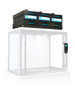
For more than 2 decades, Erlab has delivered thousands of PCR workstations. The most recent models are dedicated to 2 types of situations:
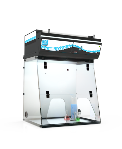
CaptairBio 320 is recommended when pre-PCR is performed in a cleanroom. The CaptairBio 320 Smart is a workstation where UV contamination is optimized to avoid cross-contamination. It features an 18W UV bulb at 254 nm that delivers a minimum of 0.08 jJ/s/cm2 at the level of the worktop.

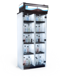
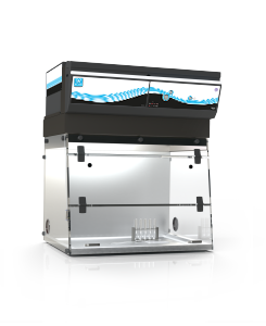
CaptairBio 321 and 391, are 2 workstations that prevent both cross-contamination and environmental contamination of samples. They can both embed HEPA or ULPA filters and are equipped with high performance 254 nm LED 900/950 lux UV bulbs.
